Sunday, January 29, 2017

Trump: The only thing we have to fear

After shocking executive actions from the White House, fear is gripping the world. The new president Trump has unleashed immigration bans, a plan to build a wall across the Mexican border, plans to cut Obamacare and more. Fear comprises our most primal emotion and we talk about it in our book series, Healing the Brain

(Book excerpt.) New York University neuroscientist Joseph LeDoux, Ph.D., and other neuroscientists have begun to examine the way the brain shapes our experience—and our memories—to generate the varied repertoire of human emotions. Specifically, as Dr. LeDoux explains, he chose to begin his inquiry by examining an emotion that is common to all living creatures: fear.

Wistar rat[edit]
Wikimedia.com
Mice serve researchers well as animal models. These very distant relatives possess well over 90 per cent of the same genes as humans.

Years of research by many workers have given us extensive knowledge of the neural pathways involved in processing acoustic information, which is an excellent starting point for examining the neurological foundations of fear. The natural flow of auditory information—the way you hear music, speech, or anything else—is that the sound comes into the ear, enters the brain, goes up to a region called the auditory midbrain, then to the auditory thalamus, and ultimately to the auditory cortex. Thus, in the auditory pathway, as in other sensory systems, the cortex is the highest level of processing.

So the first question we asked when we began these studies of the fear system was: Does the sound have to go all the way to the auditory cortex in order for the rat to learn that the sound paired with the shock is dangerous? When we made lesions in the auditory cortex, we found that the animal could still make the association between the sound and the shock, and would still react with fear behavior to the sound alone. Since information from all our senses is processed in the cortex—which ultimately allows us to become conscious of seeing the predator or hearing the sound—the fact that the cortex didn’t seem to be necessary to fear conditioning was both intriguing and mystifying. We wanted to understand how something as important as the emotion of fear could be mediated by the brain if it wasn’t going into the cortex, where all the higher processes occur.

Some other area or areas of the brain must receive information from the thalamus and establish memories about experiences that stimulate a fear response.

So we next made lesions in the auditory thalamus and then in the auditory midbrain. The midbrain supplies the major sensory input to the thalamus, which in turn supplies the major sensory input to the cortex. What we found was that lesions in either of these subcortical areas completely eliminated the rat’s susceptibility to fear conditioning. If the lesions were made in an unconditioned rat, the animal could not learn to make the association between sound and shock, and if the lesions were made on a rat that had already been conditioned to fear the sound, it would no longer react to the sound. But if the stimulus didn’t have to reach the cortex, where was it going from the thalamus?

Some other area or areas of the brain must receive information from the thalamus and establish memories about experiences that stimulate a fear response. To find out, we made a tracer injection in the auditory thalamus (the part of the thalamus that processes sounds) and found that some cells in this structure projected axons into the amygdala. This is key, because the amygdala has for many years been known to be important in emotional responses. So it appeared that information went to the amygdala from the thalamus without going to the neocortex. We then did experiments with rats that had amygdala lesions, measuring freezing and blood pressure responses elicited by the sound after conditioning. We found that the amygdala lesion prevented conditioning from taking place. In fact, the responses are very similar to those of unconditioned animals that hear the sound for the first time, without getting the shock. So the amygdala is critical to this pathway.

It receives information about the outside world directly from the thalamus, and immediately sets in motion a variety of bodily responses. We call this thalamo-amygdala pathway the low road because it’s not taking advantage of all of the higher-level information processing that occurs in the neocortex, which also communicates with the amygdala.


.

Thursday, January 26, 2017

6 Books for A Buck to Fight Trump

Project Hope is sponsored by A Thousand Moms, which supports gay youth in foster care. Our campaign is to enlighten politicians, teachers, clergy, parents--any decision maker with facts and compassion about those in our country enduring extreme stress--now more than ever.

Health is controlled in large part by our brain. Think of addiction, immune diseases, birth defects.

Buy a e-version of our books and share them. Or take a look then buy the printed book on Amazon and give it to someone you think should be informed.

6 brain books for a buck

Our well-received series on the brain is now available as e-books (like Kindle). For only a $1.00 donation to www.AThousandMoms.org

Each book is 172-192 pages, illustrated with an informative glossary. AND, each book is written at a layman's level for easy understanding. Inquire about our 60-minute documentary.

Thank you!

Donate and click the book to download the e-pub.


Healing the Brain: Stress, Trauma and LGBT/Q Youth

Healing the Brain: Stress, Trauma and Development


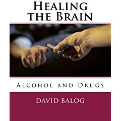
Healing the Brain: Alcohol and Drugs

Healing the Brain: Domestic Violence and TBI
Understanding  the Brain: Fetal Alcohol Spectrum Disorders
Healing the Brain: Memory
     For only a $1.00 donation to www.AThousandMoms.org    Thank you!

In pregnancy, not one drink is safe


Fetal Alcohol Spectrum Disorders describes the large number of developmental disabilities accountable to drinking alcohol while pregnant. Fetal Alcohol Syndrome is the largest single, preventable cause of developmental disabilities. In our latest Healing the Brain book, we focus on this huge problem for children--a life-long matter--and for parents and for the foster care system, where many of these children come to live.

From the book:
Fetal alcohol spectrum disorders (FASDs) are serious, lifelong birth defects and developmental disabilities caused by prenatal alcohol exposure. They are 100% preventable. Still, a surprisingly large number of children are born with FASDs each year. FASDs are a public health problem we must face. The U.S. Surgeon General has stated clearly that no amount of alcohol consumption can be considered safe for a pregnant woman and that alcohol can damage a fetus at any stage of pregnancy (Office of the Surgeon General, 2005). Yet, recent U.S. surveys reveal that approximately 12% of pregnant women still drink alcohol (CDC, 2004; SAMHSA, 2007). This means 1 in 8 fetuses are exposed to alcohol and placed at risk for FASDs. Maternal alcohol use is a growing worldwide phenomenon. It affects children and families of all ethnicities in all societies. Important international collaborative research is beginning to describe the alarming scope of this problem. While community and professional awareness of FASDs have increased, expanded awareness and informed action are sorely needed. 

FASDs are considered both medical conditions and developmental disabilities. 

FASDs cause a range of lasting medical and developmental problems and result in economic losses of billions of dollars. FASDs can also mean long-standing suffering for families. FASDs are considered both medical conditions and developmental disabilities. They include a wide range of conditions, from subtle neurodevelopmental impairments to the full fetal alcohol syndrome (FAS).  Individuals with FASDs can have physical, mental, behavioral, and/or learning disabilities with possible lifelong implications. Research shows that individuals with FASDs often have significant, long-term deficits in functional life skills.

These deficits lead to problems with day-to-day functioning as well as health care, including birth defects and increased risk for injury, unintended pregnancy, and sexually transmitted diseases. FASDs can also be associated with mental health difficulties, disrupted school and  job experiences, trouble with the law, difficulties with independent living, substance abuse, problems with parenting, and more (Bertrand et al., 2004; Streissguth et al., 2004). The median adjusted annual cost of fetal alcohol syndrome has been estimated at $3.6 billion, but the costs associated with the entire fetal alcohol spectrum are surely much higher.
 

Tuesday, January 24, 2017

The Man Who Couldn’t Remember

Our new release, Healing the Brain: Memory gives readers insight into the memory systems in our brain. They make us who we are:

This is the power of memory: a system that captures pictures, smells, sounds, events, directions--endless amounts of information every day--and then seconds or decades later calls it up for us. Memories--what we've learned and what we've done--in a large sense make us who we are. To appreciate this, think if your ability to form any new memories were suddenly cut off.

Who would you be? By studying people who've lost their memories, scientists have learned enormous amounts about how learning and memory work in healthy brains. And what they used to think was relatively straightforward they've since found is fascinatingly complex, thanks in large part to one man.


hm.jpg
Nova/PBS
The man known as H.M. made perhaps the most important contribution to the study of memory after an operation left him unable to form new long-term memories.

In 1953, radical, experimental brain surgery was used on a patient with severe epilepsy. The operation on "H.M." worked, but left him with almost no long-term memory. His case has helped scientists understand much more about the brain. For decades, the Connecticut native, known only by his initials, H.M., was the most famous patient in the study of memory and the human brain. He was a research participant for 53 years, first at the Hartford Hospital with William Beecher Scoville and Brenda Milner, then at the Montreal Neurological Institute, and after 1964 at MIT and Massachusetts General Hospital. At H.M.’s death in 2008, his full name was revealed: Henry Gustav Molaison.

The horrendous price that H.M. paid for an operation that essentially ended his seizures was a severe case of amnesia--an inability to acquire new memories, to commit to memory even the simplest events of his day or the world around him, and then to effectively retrieve those memories. This unintentional experiment, never repeated, showed that the hippocampus and medial temporal lobes are where the brain converts short-term memory into long-term memory.
       
H.M. could remember everything that happened prior to the operation. He could remember the trauma of his childhood. He could remember going to elementary school, to high school, working in the assembly plant. What he couldn't do was hold on to new information for more than a few minutes.

Monday, January 23, 2017

Trump exploits and benefits from fear, our most powerful emotion

Volumes will be written about how Donald Trump sits in the White House despite low poll numbers, and a historically low popular vote.

One element of power, examined from Machiavelli on down, is the exploitation of fear, or the acquiescence to it. Trump is the new Teflon president, fast outpacing Ronald Reagan. The more he is critiqued for obvious lies, the more support he gains, irregardless of public polls. He sits in the White House despite losing the popular vote in historic proportions.

In our book, Healing the Brain, we look at fear, the most powerful emotion of all mammals. It can be said that Trump took the low road to the White House.

Neuroscientist Joseph LeDoux, in his landmark book The Emotional Brain, looks closely at the irrational mechanisms of the fear response:

Years of research by many workers have given us extensive knowledge of the neural pathways involved in processing acoustic information, which is an excellent starting point for examining the neurological foundations of fear. The natural flow of auditory information—the way you hear music, speech, or anything else—is that the sound comes into the ear, enters the brain, goes up to a region called the auditory midbrain, then to the auditory thalamus, and ultimately to the auditory cortex. Thus, in the auditory pathway, as in other sensory systems, the cortex is the highest level of processing.

So the first question we asked when we began these studies of the fear system was: Does the sound have to go all the way to the auditory cortex in order for the rat to learn that the sound paired with the shock is dangerous? When we made lesions in the auditory cortex, we found that the animal could still make the association between the sound and the shock, and would still react with fear behavior to the sound alone. Since information from all our senses is processed in the cortex—which ultimately allows us to become conscious of seeing the predator or hearing the sound—the fact that the cortex didn’t seem to be necessary to fear conditioning was both intriguing and mystifying. We wanted to understand how something as important as the emotion of fear could be mediated by the brain if it wasn’t going into the cortex, where all the higher processes occur.

Some other area or areas of the brain must receive information from the thalamus and establish memories about experiences that stimulate a fear response.

So we next made lesions in the auditory thalamus and then in the auditory midbrain. The midbrain supplies the major sensory input to the thalamus, which in turn supplies the major sensory input to the cortex. What we found was that lesions in either of these subcortical areas completely eliminated the rat’s susceptibility to fear conditioning. If the lesions were made in an unconditioned rat, the animal could not learn to make the association between sound and shock, and if the lesions were made on a rat that had already been conditioned to fear the sound, it would no longer react to the sound. But if the stimulus didn’t have to reach the cortex, where was it going from the thalamus?

Some other area or areas of the brain must receive information from the thalamus and establish memories about experiences that stimulate a fear response. To find out, we made a tracer injection in the auditory thalamus (the part of the thalamus that processes sounds) and found that some cells in this structure projected axons into the amygdala. This is key, because the amygdala has for many years been known to be important in emotional responses. So it appeared that information went to the amygdala from the thalamus without going to the neocortex. We then did experiments with rats that had amygdala lesions, measuring freezing and blood pressure responses elicited by the sound after conditioning. We found that the amygdala lesion prevented conditioning from taking place. In fact, the responses are very similar to those of unconditioned animals that hear the sound for the first time, without getting the shock. So the amygdala is critical to this pathway.

It receives information about the outside world directly from the thalamus, and immediately sets in motion a variety of bodily responses. We call this thalamo-amygdala pathway the low road because it’s not taking advantage of all of the higher-level information processing that occurs in the neocortex, which also communicates with the amygdala.


Saturday, January 14, 2017

Will I get Alzheimer's?

Alzheimer's is a scary word. What is the reality, the science, the hope? Here's a sample from Healing the Brain: Memory. Click the link to download the entire Healing the Brain Series. Six books for a buck.


Alzheimer's Disease--Facts and Treatments

Alzheimer’s disease is an irreversible, progressive brain disorder that slowly destroys memory and thinking skills, and eventually the ability to carry out the simplest tasks. In most people with Alzheimer’s, symptoms first appear in their mid-60s. Estimates vary, but experts suggest that more than 5 million Americans may have Alzheimer’s.


... Alzheimer's Disease - Stem ...
PlacidWay.com
While there is no cure for Alzheimer’s disease, researchers are making advances on treatments that delay the onset and progress of the disease.

Alzheimer's disease is currently ranked as the sixth leading cause of death in the United States, but recent estimates indicate that the disorder may rank third, just behind heart disease and cancer, as a cause of death for older people.


Alzheimer’s is the most common cause of dementia among older adults. Dementia is the loss of cognitive functioning—thinking, remembering, and reasoning—and behavioral abilities to such an extent that it interferes with a person’s daily life and activities. Dementia ranges in severity from the mildest stage, when it is just beginning to affect a person’s functioning, to the most severe stage, when the person must depend completely on others for basic activities of daily living.


The causes of dementia can vary, depending on the types of brain changes that may be taking place. Other dementias include Lewy body dementia, frontotemporal disorders, and vascular dementia. It is common for people to have mixed dementia—a combination of two or more disorders, at least one of which is dementia. For example, some people have both Alzheimer's disease and vascular dementia.


In 1906, Dr. Alois Alzheimer noticed changes in the brain tissue of a woman who had died of an unusual mental illness. Her symptoms included memory loss, language problems, and unpredictable behavior. After she died, he examined her brain and found many abnormal clumps (now called amyloid plaques) and tangled bundles of fibers (now called neurofibrillary, or tau, tangles).


These plaques and  tangles in the brain are still considered some of the main features of Alzheimer’s disease. Another feature is the loss of connections between nerve cells (neurons) in the brain. Neurons transmit messages between different parts of the brain, and from the brain to muscles and organs in the body.

Where were you on 9/11?

Here's an excerpt from Healing the Brain: Memory. Learn more about the brain in this special offer: 6 brain books for a buck.


Emotions and “flashbulb” memories.
Your emotional state when an event occurs can greatly influence your memory of it. Thus, if an event is very upsetting, you will form an especially vivid memory of it. For example, many people remember where they were when they learned about President Kennedy’s assassination, or about the attacks of September 11, 2001. The processing of emotionally-charged events in memory involves norepinephrine, a neurotransmitter that is released in larger amounts when we are excited or tense. As Voltaire put it, that which touches the heart is engraved in the memory.

Location, light, sounds, smells...in short, the entire context in which the memorizing takes place is recorded along with the information being memorized. Our memory systems are thus contextual. Consequently, when you have trouble remembering a particular fact, you may be able to retrieve it by recollecting where you learned it or the book from which you learned it. Was there a picture on that page? Was the information toward the top of the page, or the bottom? Such items are called “recall indexes”. And because you always memorize the context along with the information that you are learning, by recalling this context you can very often, by a series of associations, recall the information itself.